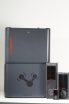
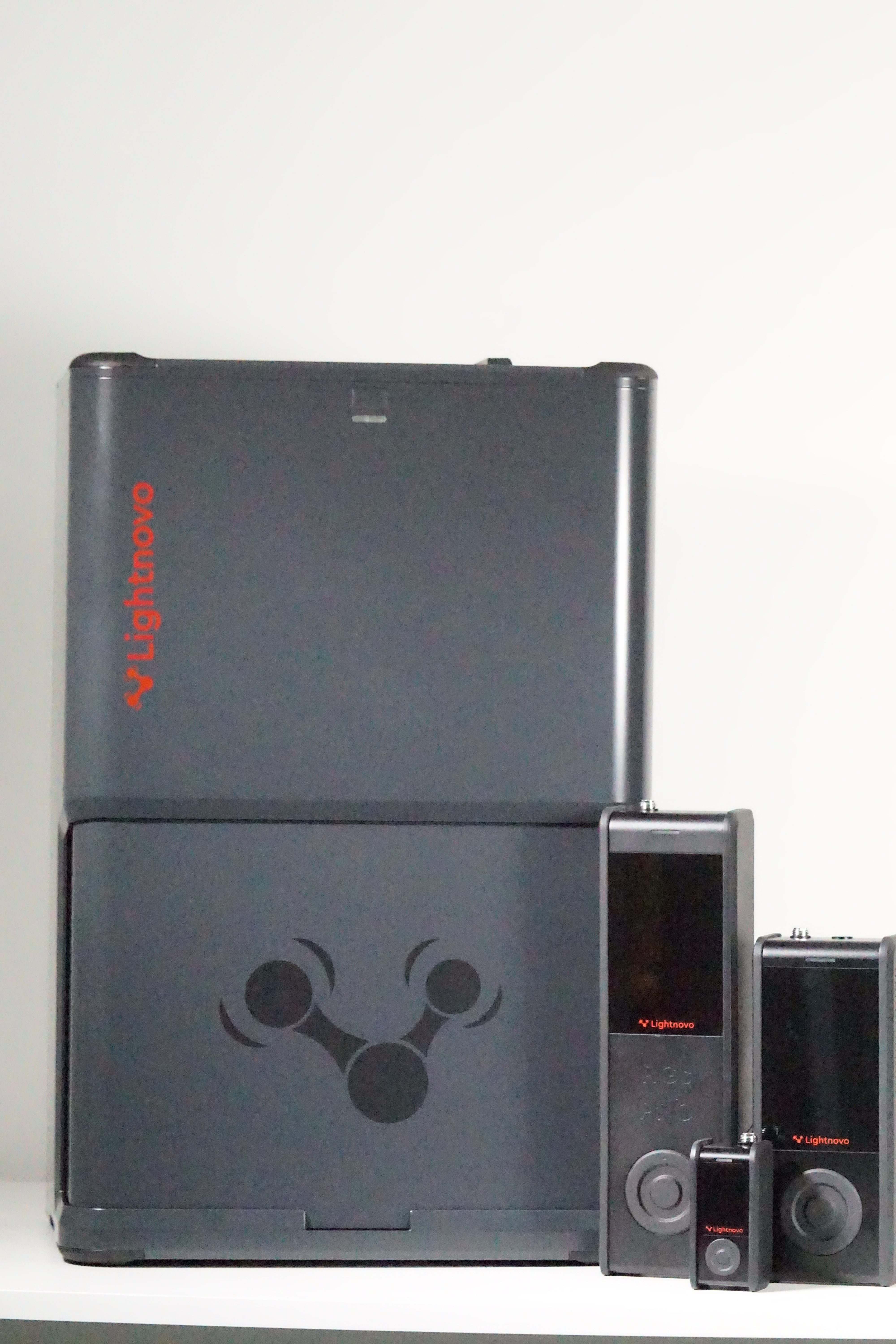
RG Microscope
Mit ihrem erstklassigem Research Grate (RG) Microscope der 3. Generation liefert Lightnovo ein konfokales Raman-Mikroskop für die moderne Forschung. Das RG Microscope ist die Antwort auf die leistungsstarke und anspruchsvolle Raman-Mikroskopie, bei der eine hohe spektrale und räumliche Auflösungen, ein erweiterter Spektralbereich und eine außergewöhnliche Laserstabilität benötigt wird.
Zu den wichtigsten Eigenschaften des RG Microscopes zählen das modulare und kompakte Design, die hohe spektrale Auflösung, der große „Mapping“ Bereich, die Auswahl an verschiedenen Lasern durch die vielfältigen Raman Geräte und das rasche Wechseln von der aufrechten- zur invertierten Mikroskopie ohne Umbau des Mikroskops.
Eigenschaften
Die Besonderheit des RG Microscopes ist, dass jedes Lightnovo Raman Spektrometer der 3. Generation verwendet werden kann: miniRaman und miniRaman Pro, RG Raman und RG Raman Pro. Für das miniRaman Spectrometer muss ein Adapter angeschraubt werden, damit das Spektrometer selbstjustiert im Mikroskop ist; für das RG Spectrometer gibt es eine direkte Halterung, was ein weiteres Alignment überflüssig macht.
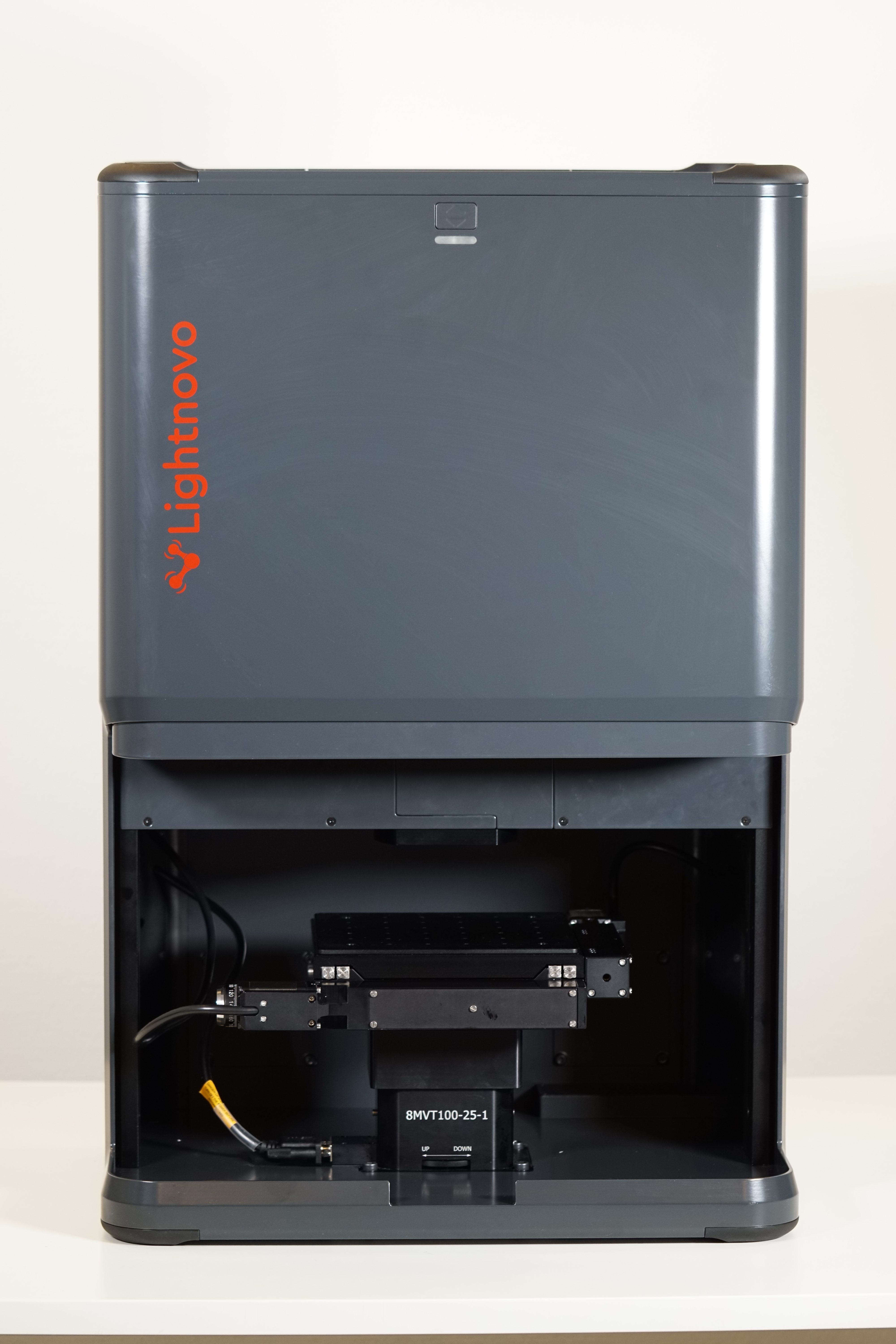
Wie beim miniRaman Spectrometer kann auch das RG Spectrometer in aufrechtem und invertiertem Mikroskopie-Modus bedient werden, einfach durch das Umdrehen des Gerätes. Dafür ist weder Neumontage noch Umbau nötig. Mit einem Gewicht von 34 bis 43 kg – abhängig von der Konfiguration – und einer Größe von 360x450x685 mm (LxBxH), kann das Gerät mit etwas Geschick umgedreht werden.
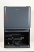
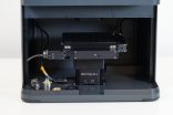
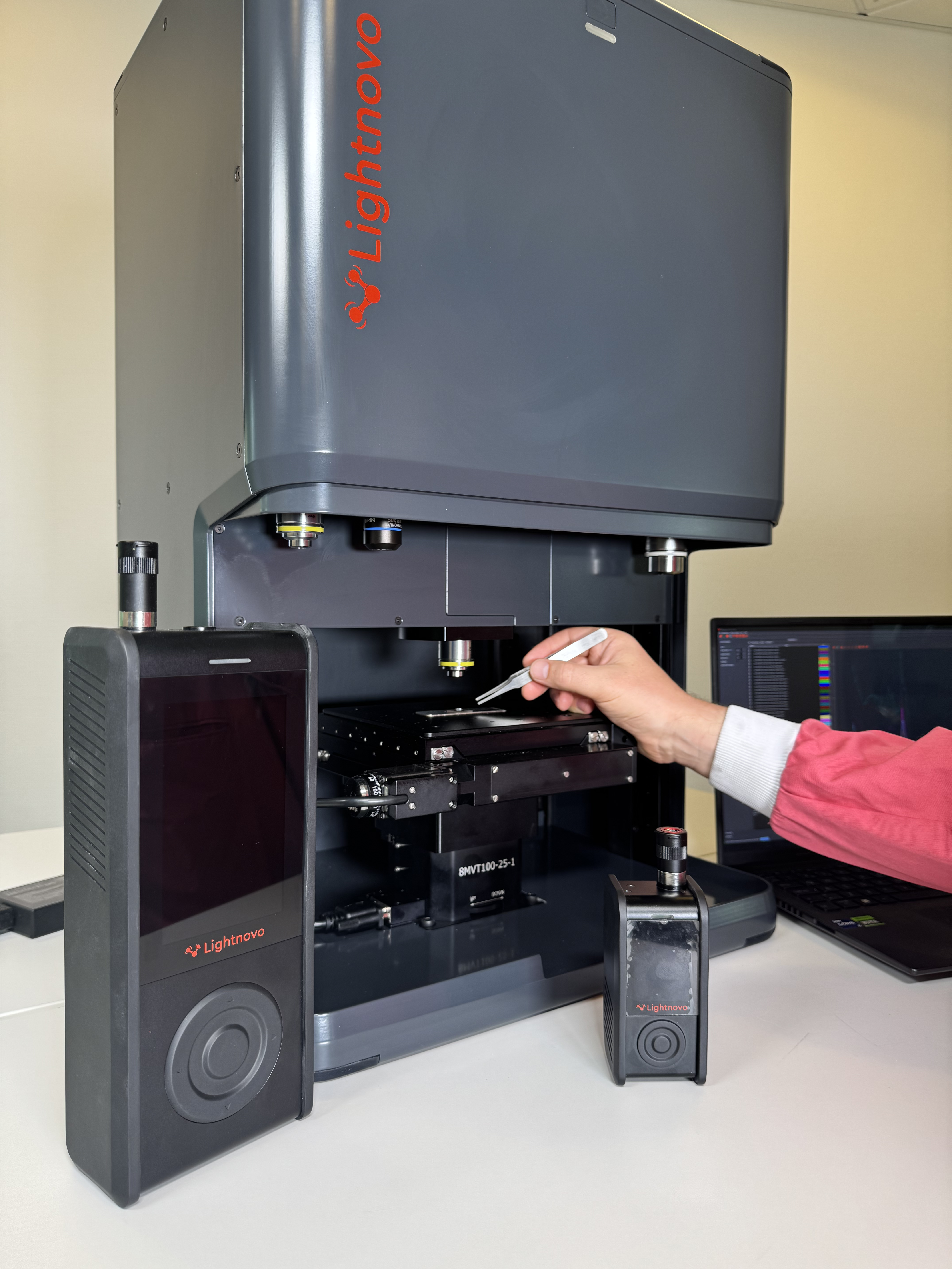
Ausgestattet mit einem Auflichtmikroskop (reflected light microscope) mit separatem Kamerasensor können mit dem RG Microscope gleichzeitig optische Parameter (Größe, Morphologie) und ein Raman-Spektrum (chemische und strukturelle Analyse) erfasst werden, d.h. der Laserspot kann im Mikroskop-Bild beobachtet werden und liefert Informationen über den Ort des Raman-Mappings.
Das RG Microscope verfügt über eine hohe spektrale und räumliche Auflösung: die laterale Auflösung beträgt 200 – 420 nm und die axiale Auflösung 700 – 2400 nm (Werte sind abhängig vom verwendeten Spektrometer), die lateralen Schrittgröße 100 nm.
Das RG Microsope wird der Laserklasse 1 zugeordnet; beim Öffnen der automatischen Türe wird der Laser direkt ausgeschaltet. Des Weiteren kann das Gehäuse als Inkubator, beispielsweise für Zellen und Bakterien, verwendet werden.
Anstelle eines sehr voluminösen Objektivrevolvers, wird beim RG Microscope auf magnetische Haltepunkte für zusätzliche Objektive gesetzt. Mit dem magnetischen Adapter können die Objektive somit einfach getauscht werden. Zusätzlich bleibt die Stage automatisch stehen, falls eine Kraft von mehr als 5kg auf das Objektiv wirkt. Dieser Mechanismus verhindert, dass die Probe beschädigt wird .


Software
Die Software Miraspec kann am PC (Windows 11) und/oder dem Smartphone (Android) verwendet werden. Sie bietet neben den Funktionen, die für das Ansteuern des Raman Gerätes und der Spektren-Erfassung benötig werden (siehe RG Spectrometer), auch die Möglichkeit, mikroskopische Informationen zu erfassen. So kann gleichzeitig ein Bild aufgenommen und der Laserspot dargestellt, oder eine Panorama-Probenabbildung durchgeführt werden.
Beim sogenannten „Mapping“ ist eine X-, Y-, und Z-Raumabbildung (spatial mapping), eine Zeitabbildung (time mapping) und eine Krümmungskompensation möglich, außerdem das Messen der Peak-Intensitäts-Karte und Peak-Flächen-Karte unter Verwendung von 2 Dimensionen (X, Y, Z, Zeit oder Raman-Verschiebung).
Zubehör
- Magnetische Objektivadapter für schnellen Wechsel
- Objektive mit Standardgewinde (RMS, M25)
- Mikroskop-Objektive mit folgender Vergrößerung: 10-fach, 20-fach, 50-fach und 100-fach
- Kundenspezifische Mikroskop-Objektive auf Anfrage
- Objektivabdeckungen und Mikroskop-Objektträgerhalter
- 96-Well-Plattenhalter
Anwendungsbereiche
Das RG Microscope ist für den Einsatz in Universitäten, Forschung & Entwicklung und Industrie geeignet und findet Anwendungen in den Bereichen Biowissenschaften, Nanotechnologie, Materialwissenschaften und industrielle Analyse.
- Biowissenschaften und Gesundheit: Biowissenschaften, Pharmazeutika, Hautdiagnostik, Kosmetika
- Materialien und Nanotechnologie: Polymere, Nanomaterialien, Halbleiter, oberflächenverstärkte Raman-Streuung (SERS)
- Industrielle und chemische Analyse: Chemikalien, Geologie, Forensik
- Qualitätskontrolle und Authentifizierung: Alkoholqualität, Erkennung von gefälschten Produkten
Technical specifications
| Feature versus model | RG microscopy platform motorized | RG microscopy platform manual | RG microscopy platform focusing stage |
|---|---|---|---|
| Compatible spectrometers* | miniRaman spectrometers, miniRaman Pro spectrometers, RG spectrometers, RG Pro spectrometers | ||
| Lateral resolution** | 200 - 420 nm | ||
| Axial resolution or confocality** | 700 - 2400 nm | ||
| White light microscopy | Real-time camera visualization of the sample reflected with simultaneous visualization of laser spot and Raman acquisition | ||
| Microscopy configuration | up-right and inverted | ||
| Mapping range in XYZ | 102 x 102 x 25 mm | 15 x 15 x 15 mm | |
| Minimum step size | 100 nm | 5 µm | |
** Resolution depends on the laser wavelength and spectrometer used. Values represent the use of RG spectrometers, all wavelengths with microscope objective NA=0,95, 100x magnification