RG Microscope
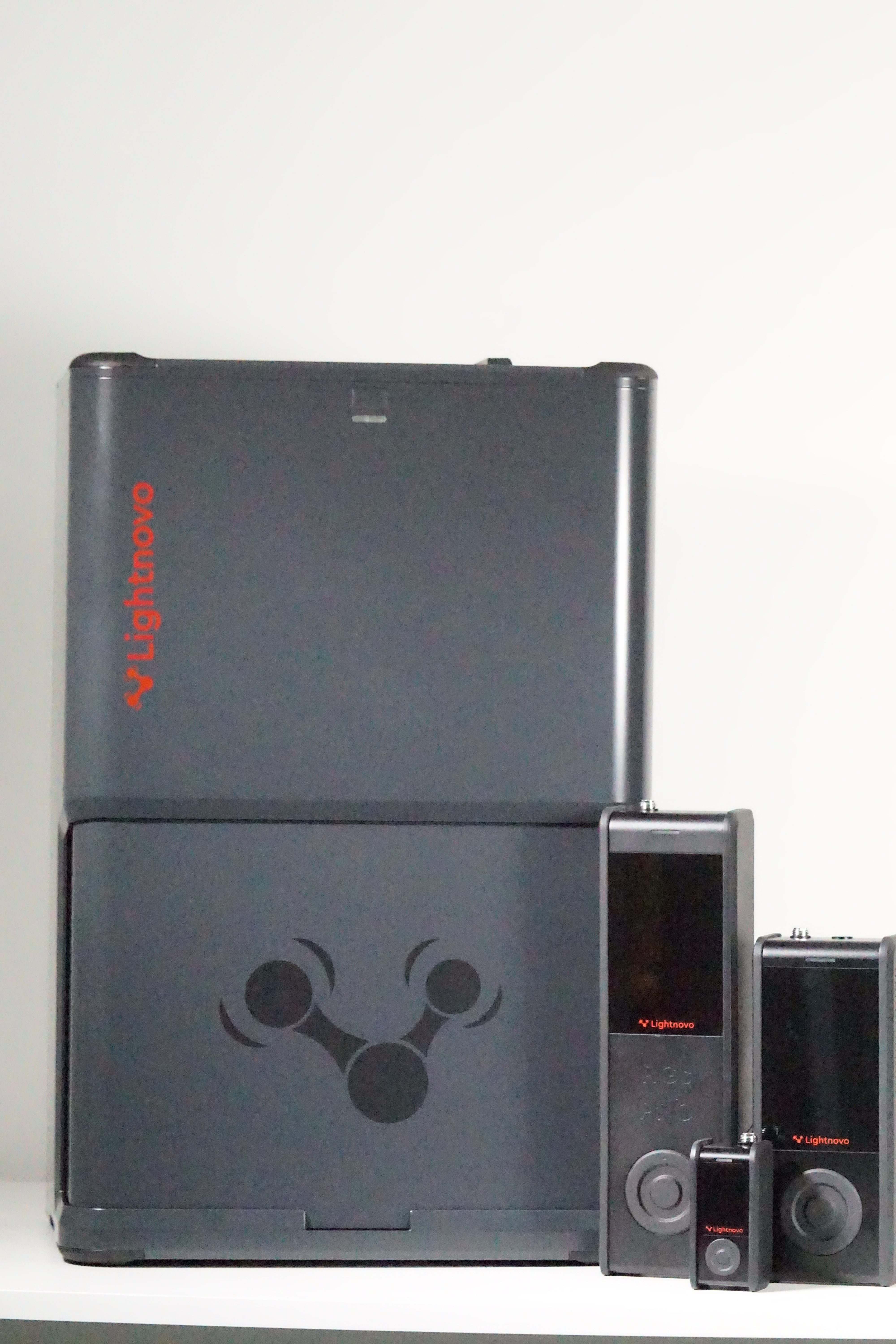
With its first-class third-generation Research Grate (RG) Microscope, Lightnovo delivers a confocal Raman microscope for modern research. The RG Microscope is the answer to powerful and demanding Raman microscopy, which requires high spectral and spatial resolutions, an extended spectral range, and exceptional laser stability.
The most important features of the RG Microscope include its modular and compact design, high spectral resolution, large mapping range, selection of different lasers thanks to the wide range of Raman devices, and quick switching from upright to inverted microscopy without having to modify the microscope.
Features
The special feature of the RG Microscope is that any 3rd generation Lightnovo Raman spectrometer can be used: miniRaman and miniRaman Pro, RG Raman and RG Raman Pro. An adapter must be screwed onto the miniRaman Spectrometer so that the spectrometer is self-aligned in the microscope; for the RG Spectrometer, there is a direct mount, which eliminates the need for further alignment.
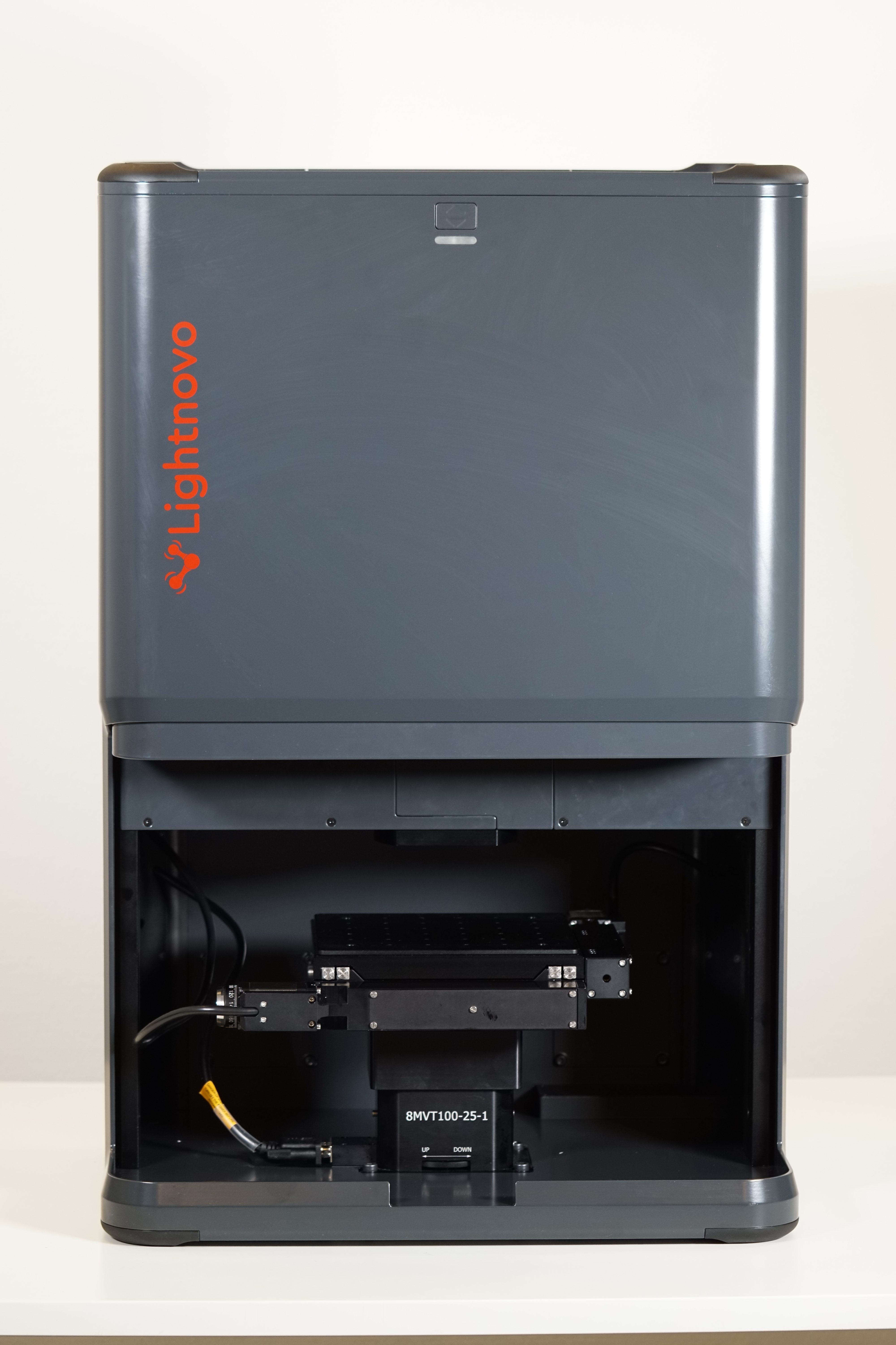
As with the miniRaman Spectrometer, the RG Spectrometer can also be operated in upright and inverted microscopy mode simply by turning the device upside down. No new installation or conversion is necessary for this. Weighing between 34 and 43 kg, depending on the configuration, and measuring 360x450x685 mm (LxWxH), the device can be turned over with a little skill.
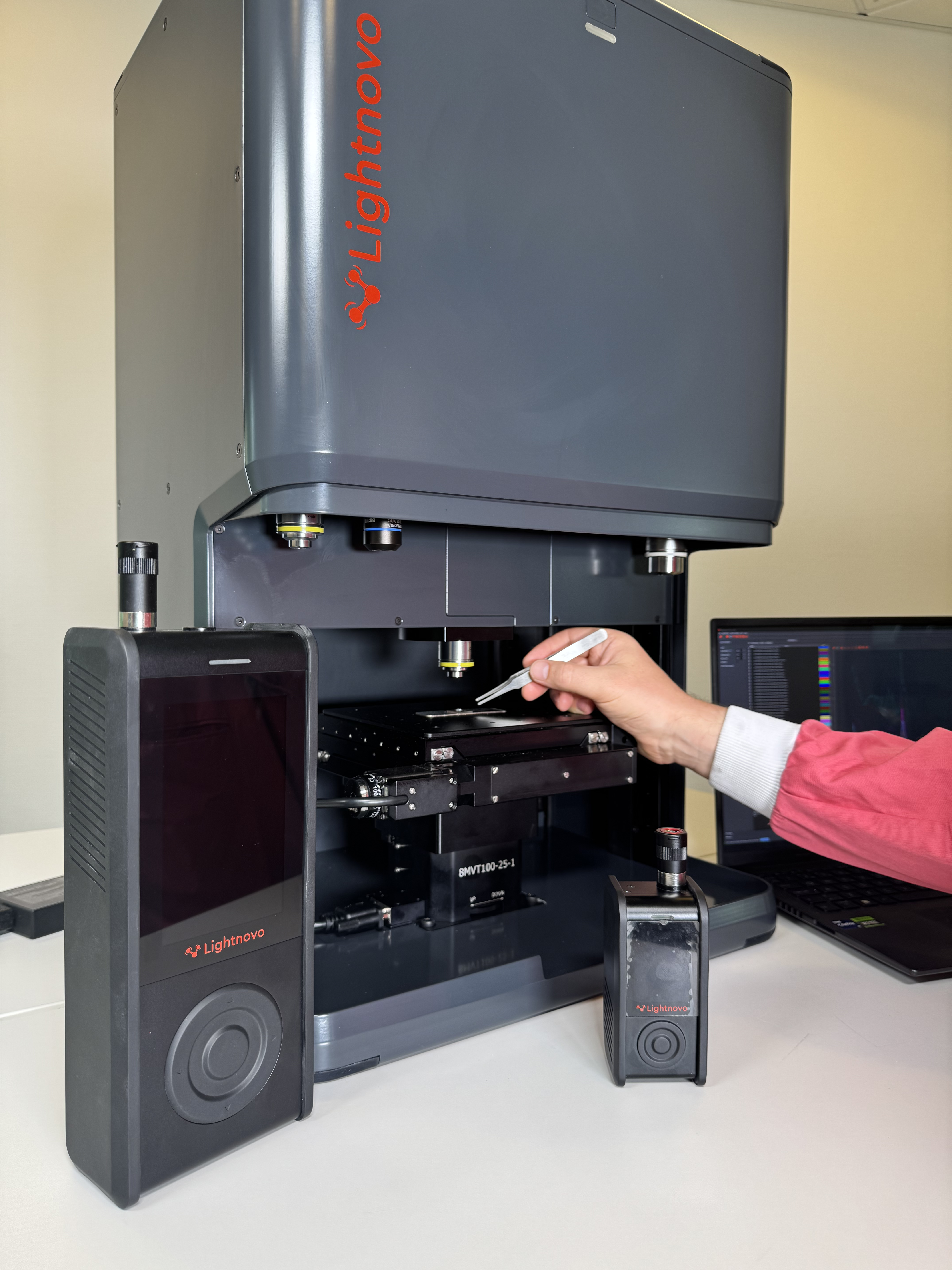
Equipped with a reflected light microscope with a separate camera sensor, the RG Microscope can simultaneously capture optical parameters (size, morphology) and a Raman spectrum (chemical and structural analysis), i.e., the laser spot can be observed in the microscope image and provides information about the location of the Raman mapping.
The RG Microscope has high spectral and spatial resolution: the lateral resolution is 200 – 420 nm and the axial resolution is 700 – 2400 nm (values depend on the spectrometer used), with a lateral step size of 100 nm.
The RG Microscope is classified as a Class 1 laser; the laser is switched off immediately when the automatic door is opened. The housing can also be used as an incubator, for example for cells and bacteria.
Instead of a very bulky objective revolver, the RG Microscope uses magnetic mounting points for additional objectives. The magnetic adapter makes it easy to swap objectives. In addition, the stage automatically stops if a force of more than 5 kg is applied to the objective. This mechanism prevents damage to the sample.


Software
The Miraspec software can be used on a PC (Windows 11) and/or smartphone (Android). In addition to the functions required for controlling the Raman device and spectrum acquisition (see RG Spectrometer), it also offers the option of capturing microscopic information. This allows an image to be captured and the laser spot to be displayed at the same time, or a panoramic sample image to be taken.
So-called “mapping” enables X, Y, and Z spatial mapping, time mapping, and curvature compensation, as well as the measurement of peak intensity maps and peak area maps using two dimensions (X, Y, Z, time, or Raman shift).
Accessories
- Magnetic lens adapters for quick changes
- Lenses with standard thread (RMS, M25)
- Microscope objectives with the following magnifications: 10x, 20x, 50x, and 100x
- Custom microscope lenses available on request
- Lens covers and microscope slide holders
- 96-well plate holder
Areas of application
The RG Microscope is suitable for use in universities, research and development, and industry, and has applications in the fields of life sciences, nanotechnology, materials science, and industrial analysis.
- Life sciences and health: Life sciences, pharmaceuticals, skin diagnostics, cosmetics
- Materials and nanotechnology: polymers, nanomaterials, semiconductors, surface-enhanced Raman scattering (SERS)
- Industrial and chemical analysis: chemicals, geology, forensics
- Quality control and authentication: alcohol quality, detection of counterfeit products
Technical specifications
| Feature versus model | RG microscopy platform motorized | RG microscopy platform manual | RG microscopy platform focusing stage |
|---|---|---|---|
| Compatible spectrometers* | miniRaman spectrometers, miniRaman Pro spectrometers, RG spectrometers, RG Pro spectrometers | ||
| Lateral resolution** | 200 - 420 nm | ||
| Axial resolution or confocality** | 700 - 2400 nm | ||
| White light microscopy | Real-time camera visualization of the sample reflected with simultaneous visualization of laser spot and Raman acquisition | ||
| Microscopy configuration | up-right and inverted | ||
| Mapping range in XYZ | 102 x 102 x 25 mm | 15 x 15 x 15 mm | |
| Minimum step size | 100 nm | 5 µm | |
** Resolution depends on the laser wavelength and spectrometer used. Values represent the use of RG spectrometers, all wavelengths with microscope objective NA=0,95, 100x magnification